Welcome to Bayesil


You can now download results in nmrML format. Upload options for this format are coming soon. Please stay tuned!


Home spectra

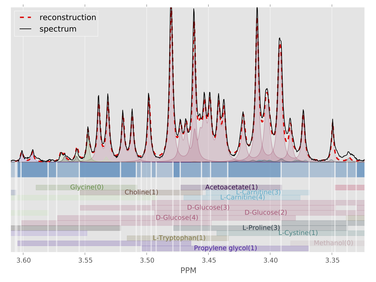
Bayesil is a web system that automatically identifies and quantifies metabolites using 1D 1H NMR spectra of ultra-filtered plasma, serum or cerebrospinal fluid. The NMR spectra must be collected in a standardized fashion (see How To Collect NMR Spectra for Bayesil) for Bayesil to perform optimally. Bayesil first performs all spectral processing steps, including Fourier transformation, phasing, solvent filtering, chemical shift referencing, baseline correction and reference line shape convolution automatically. It then deconvolutes the resulting NMR spectrum using a reference spectral library, which here contains the signatures of more than 60 metabolites (see here for a list). This deconvolution process determines both the identity and quantity of the compounds in the biofluid mixture. Extensive testing shows that Bayesil meets or exceeds the performance of highly trained human experts.

Citing Bayesil:

Ravanbakhsh S, Liu P, Bjorndahl TC, Mandal R, Grant JR, Wilson M, Eisner R, Sinelnikov I, Hu X, Luchinat C, Greiner R, Wishart DS. (2015) Accurate, Fully-Automated NMR Spectral Profiling for Metabolomics. PLoS ONE 10(5): e0124219.

Nmr add

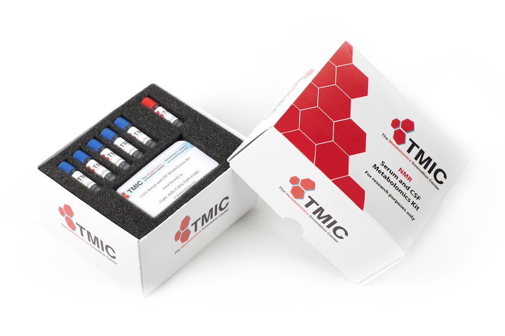
NMR Metabolomics Analysis Kit

Serum and CSF

• For preparation of 50 serum or CSF samples
• Processing time: 2 min/sample
• Access to Bayesil batch processing feature
• Process and analyse samples for as low as $10/sample


More info hereClick to order

Bayesil Spectral Analysis

Instructions

To analyze a 1D 1H NMR spectrum with Bayesil you must provide information on the biofluid being analyzed, the concentration of the reference standard, the spectrometer frequency and the 1D NMR spectral file.

Run one of our examples:
Example 1

Biological Serum Varian 500 MHz

Example 2

Biological CSF Varian 500 MHz

Example 3

Biological Serum Bruker 600 MHz

Or submit your own mixture:

  1. Bayesil works with any mammalian serum, plasma or CSF. For other biofluids use Custom to define a spectral library for profiling.

  2. Provide the known concentration of the Chemical Shift reference in your sample. This will be used to quantify the profiled metabolites.
    µM

  3. Provide the frequency of the NMR spectrometer used to collect the spectrum.

  4. Please ensure your spectrum has been collected using the protocol described here. The uploaded file must be a ZIP compressed FID folder (Agilent/Varian or Bruker).

  5. Fast Profile is faster but less acurate.